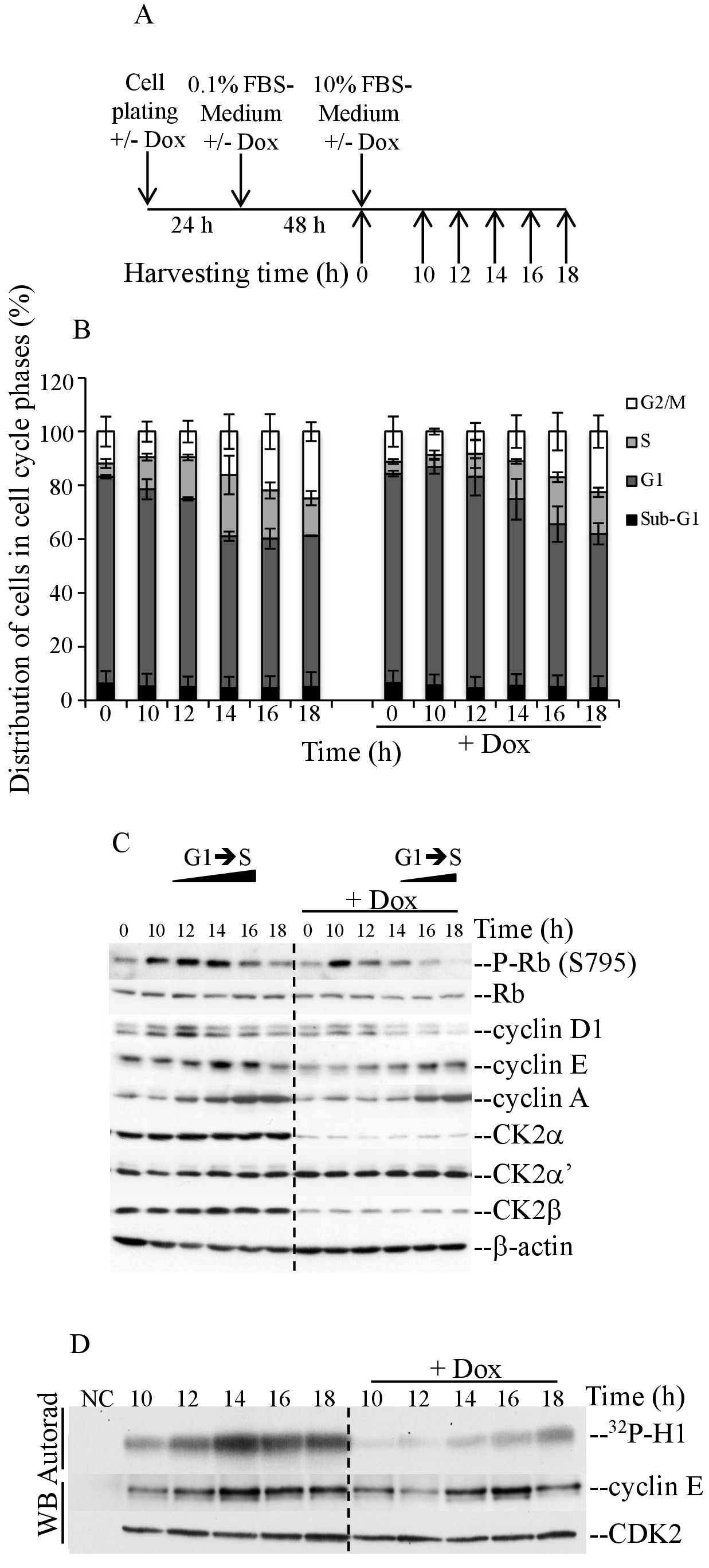
Fig. 2. Silencing of protein kinase CK2α results in marked delay in cell progression through G1 and inhibition of cyclin E-CDK2 activity. (A, B) 24 h after plating, cells were starved in growth medium containing 0.1% fetal bovine serum for 48 hours in the presence or absence of doxycycline as indicated in (A). Cells were collected at the indicated time points after adding full growth medium and subsequently analyzed by FACS as shown in (B). (C) Cells were treated as indicated in (A) and then harvested for protein expression analysis. Whole cell lysates were examined by western blot employing antibodies against the indicated proteins. (D) Whole lysate from cells treated as described in (A) was subjected to immunoprecipitation in the presence of either control serum (NC) or mouse monoclonal anti-CDK2 antibody. Immunoprecipitates were subjected to phosphorylation assay employing histone H1 as substrate target. Phosphorylation of histone H1 was revealed by autoradiography (Autorad). Immunoprecipitates were analyzed by western blot using the indicated antibodies (WB). Experiments were repeated at least three times obtaining similar results.